General dentists provide services related to the general maintenance of oral hygiene and dental health
Dental radiography
There are two categories of radiography:
2D X-rays
3D X-rays
2D X-rays include:
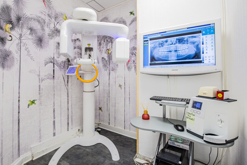
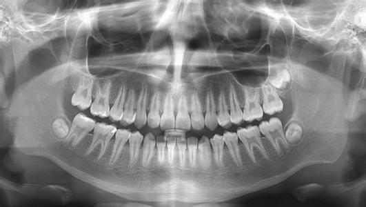
Panoramic radiography : it is performed within the office, usually in a dedicated room, and will provide information and insight into all of the teeth in the mouth, but also on the teeth that did not come out (like the wisdom teeth or the erupting tooth germs with children). By the global vision it offers, the level of the gums and the bone around the teeth is also visible. It is usually performed every year or two at the time of the follow-up visit, in order to compare it with an X-ray of the previous check-up and to see directly if a tooth or a restoration has deteriorated since the last check-up. This radiography, performed with the new generation of equipment used today, is low-radiation.
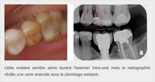
Intra-oral dental radiography : This is a small X-ray performed directly on the dentist’s chair, which will give more detailedinformation on three or four adjacent teeth. It is performed when a tooth needs to be evaluated in more detail if there is a suspicion of pathology on it, or when pain is described precisely by the patient on a specific area of the mouth. It is then compared with the dentist’s clinical examination and any other panoramic or 3D radiographs.
Profile teleradiography (or cephalometric radiography): This is an x-ray of the jawbones, skull and teeth, taken from the side, in a radiology center, on prescription from the dentist or orthodontist. It is performed during an orthodontic check-up so that the orthodontist can diagnose the situation with a global vision of the jaws and teeth, and their angulations.
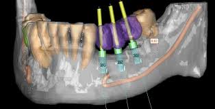
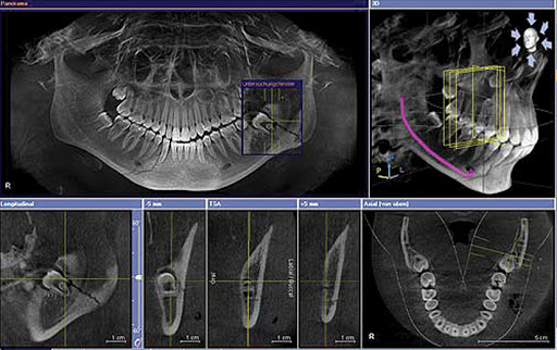
Among the 3D X-rays, we will find mainly in dentistry the dental Cone Beam (very close to the scanner, but less radiating). The Cone Beam can be performed directly in our dental office, equipped with one of the new three-dimensional radiology devices capable of giving scans and 3D information of the bones and joints of the jaw, the anatomical areas bordering the jaw (Example: Maxillary sinus), and teeth in less than 5 minutes.
The dental Cone Beam is performed in certain cases only:
Pre-implant check-up: to see the bone conditions of the jaw and plan a dental implant placement. It will also allow us to plan a pre-implant bone graft if the bone is not of sufficient quality to receive an implant
In-depth assessment of a tooth: In certain cases, the dentist can use the Cone Beam to obtain more information on a particular tooth for example in case of recurrent infection under a devitalized tooth, or following a trauma with suspicion of a crack on the root of a tooth or a fracture of the jaw. The 3D X-ray will allow the dentist to view the tooth from all angles, and also to see hundreds of slices of the inside of the tooth.